

Working with Us
AtlasXomics offers researchers flexible options for using our platform, including sending samples to our service or integrating the platform directly into their labs with our kits
Service
1. Meeting with Service lead to understand biological question and region of interest

2. Sample optimization/qualification to determine feasibility of sample or antibody target

3. Spatial analysis, sequencing and standard data report with raw data

4. Visualize processed data in our interactive shinyApp

Our team collaborates closely with the investigator to identify and interrogate their region of interest (ROI) that best addresses their biological question.
The investigator will provide samples to be qualified for spatial analysis. Our team will recommend whether the samples will generate high-quality spatial data.
Once qualified, the samples will be analyzed and a detailed data report will be provided to the customer
Tissue Requirements
•OCT embedded fresh frozen samples, stored at -80C
•Mount 7-10 μm tissue sections onto 1”x3” slides coated for tissue adhesion
•Each tissue region of interest should be centered on the slide (please communicate to AtlasXomics team your area of interest)
•One region of interest per slide (can contain multiple sections if they are within the region of interest)

Sample Preparation and Shipping Protocols
Kits
Our DBiT-seq platforms offers a comprehensive solution, equipping you with all the essential reagents and microfluidic chips required for efficient Tagmentation and spatial barcoding. Designed for easy adoption, our microfluidic chips are dust resistant and compatible with multi-channel pipettes, ensuring a hassle-free integration of our platform.
Microfluidic Chips


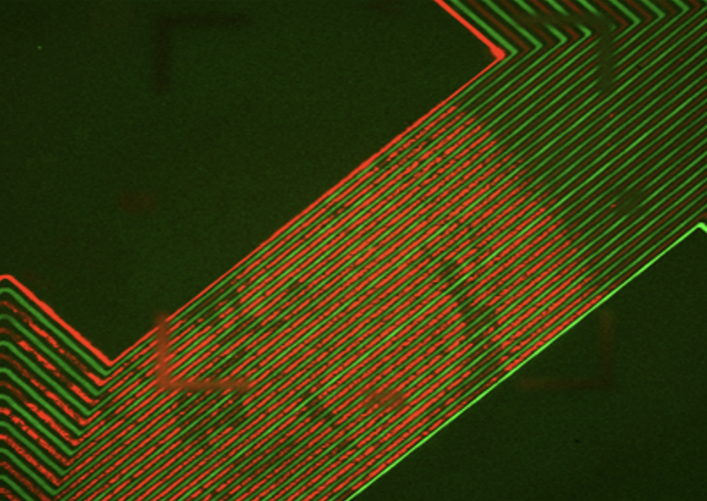
X and Y Chip Pair with spatial barcodes
Consumable reagants
Our spatial ATAC-seq kit includes: our microfluidic chips containing spatial barcodes, Tn5 Transposase loaded with our custom adapters, sequencing adapters, buffers and enzymes.
We also recommend using our Sample Optimization Kit to optimize the Tn5 reaction in different tissue types and ensure high quality reagent delivery with our microfluidics: This kit includes flow QC chips and bulk ATAC-seq reagents.
Custom Hardware
AtlasXpress

Seals our microfluidic chips to tissue mounted slide
Leveraging standard workflows
Epifluorescence scanning microscope with tiling capacity (e.g. Keyence BZ-X, EVOS M7000)
Access to Next Generation Sequencer (NGS): spatial ATAC-seq requires 200-300M reads 150 Paired-end (60-90 Gb/sample)
Standard PCR/NGS library preparation materials and equipment
Positive pressure source (optional) : Facilitates slide/tissue drying